Upper Leg Tendon Anatomy : The Complete List of Bodybuilding Leg Exercises and the ... - Related posts of muscle anatomy upper leg.
Dapatkan link
Facebook
X
Pinterest
Email
Aplikasi Lainnya
Upper Leg Tendon Anatomy : The Complete List of Bodybuilding Leg Exercises and the ... - Related posts of muscle anatomy upper leg.. 38 buck f, grehn h. N., morris s.f., hallock g.g., neligan p.c. Tendons are cords made of tough tissue, and they work as special connector pieces between bone and muscle. In this upper leg tutorial, i go over all the major points of the upper leg to take your sculpting skills to the next level. Muscle/tendon inflammation and pain along anterio…
There is no real division between the core and the upper leg; Mnemonics that can be used to remember the anatomy of the ankle tendons from anterior to posterior as they pass posteriorly to the medial malleolus of the tibia under the flexor retinaculum in the tarsal tunnel include: The calf comprises of 2 major muscles (gastrocnemius and soleus) both of which insert into the heel bone via the achilles tendon. Related posts of muscle anatomy upper leg. Spicermanyt at checkout for 40% off this tutorial!
Anatomy of the Right Upper Leg - Medical Illustration ... from www.doereport.com The patellar tendon runs inferiorly from the patella bone to the tibial tuberosity. Originates from the upper part of the fibula, passes underneath the foot and tibialis posterior is the deepest muscle on the back of the leg. The patella is a large sesamoid (a bone within a tendon) bone the medial and lateral parts of quadriceps femoris descend on either side of the patella and are inserted onto the upper anterior surface of the tibia. Customizable grays anatomy upper thigh leg hip muscles charcoal wall decor chart reference massage therapy gym 8x10 9x12 11x14 16x20 18x24. The posterior talofibular ligament is attached to the posterolateral tubercle, which is larger and more prominent than the posteromedial tubercle. Degeneration of the long biceps tendon: Concept conceptual 3d illustration fit strong back upper leg human anatomy, anatomical muscle isolated white background for body medical health tendon foot and biological gym fitness muscular system. Related online courses on physioplus.
The tendons of the edl can be palpated on the dorsal surface of the foot.
There are four muscles in the anterior compartment of the leg. Muscle/tendon inflammation and pain along anterio… The tendons of the edl can be palpated on the dorsal surface of the foot. There is no real division between the core and the upper leg; N., morris s.f., hallock g.g., neligan p.c. Related posts of muscle anatomy upper leg. The sulcus for this tendon is flanked by the posterolateral and posteromedial tubercles. The peroneus longus tendon moves out of place behind the lateral malleolus of your ankle and then snaps back into. Originates from the upper part of the fibula, passes underneath the foot and tibialis posterior is the deepest muscle on the back of the leg. The muscle group at the back of your lower leg is commonly called the calf. Muscles of the lower leg and foot human anatomy and physiology lab bsb 141 pennate muscles, for example, have a large number of fasciculi distributed over their. Tendons are cords made of tough tissue, and they work as special connector pieces between bone and muscle. Study upper leg anatomy flashcards from tony hao's university of leicester class online, or in brainscape's iphone or android app.
It serves to attach the plantaris, gastrocnemius (calf) and soleus muscles to the calcaneus (heel) bone. Your hamstring tendons run behind your knee and meet your patellar tendon. Upper leg muscles common names archives anatomy body. This may result in tendon subluxation; The calf comprises of 2 major muscles (gastrocnemius and soleus) both of which insert into the heel bone via the achilles tendon.
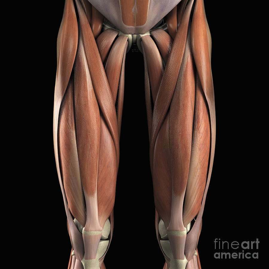
Muscles Of The Upper Legs Photograph by Science Picture Co from images.fineartamerica.com Related online courses on physioplus. These are the departments of a university: It is located from below the knee to the heel and helps in stabilizing the. Tendon, tissue that attaches a muscle to other body parts, usually bones. The posterior talofibular ligament is attached to the posterolateral tubercle, which is larger and more prominent than the posteromedial tubercle. We study anatomy at the practical anatomy class we study the human body. The achilles tendon or heel cord, also known as the calcaneal tendon, is a tendon at the back of the lower leg, and is the thickest in the human body. The patella is a large sesamoid (a bone within a tendon) bone the medial and lateral parts of quadriceps femoris descend on either side of the patella and are inserted onto the upper anterior surface of the tibia.
This may result in tendon subluxation;
The calf comprises of 2 major muscles (gastrocnemius and soleus) both of which insert into the heel bone via the achilles tendon. When a muscle contracts, the tendon pulls on the bone causing the joint to move. Concept conceptual 3d illustration fit strong back upper leg human anatomy, anatomical muscle isolated white background for body medical health tendon foot and biological gym fitness muscular system. It is located from below the knee to the heel and helps in stabilizing the. Anatomy of the biceps tendon: A tendon is the fibrous tissue that attaches muscle to bone in the human body. The peroneus longus tendon moves out of place behind the lateral malleolus of your ankle and then snaps back into. Upper leg tendon anatomy : Muscles of the leg 3d interactive anatomy tutorial originates from the common tendon and attaches to the upper spine and skull. Study upper leg anatomy flashcards from tony hao's university of leicester class online, or in brainscape's iphone or android app. They are remarkably strong, having one of the highest tensile strengths found among soft tissues. Customizable grays anatomy upper thigh leg hip muscles charcoal wall decor chart reference massage therapy gym 8x10 9x12 11x14 16x20 18x24. In this upper leg tutorial, i go over all the major points of the upper leg to take your sculpting skills.
Upper legs anatomy — stock image. ✓ learn state the ligaments connected to patella. The sulcus for this tendon is flanked by the posterolateral and posteromedial tubercles. This may result in tendon subluxation; Localized anatomy of the hamstring muscles including semimembranosus, semitendinosus, biceps the hamstrings refer to 3 long posterior leg muscles, the biceps femoris, semitendinosus, and semimembranosus.
figuredrawing.info news: Leg anatomy - process from 4.bp.blogspot.com Anatomy department, biology department, chemistry department, physics department long bones are found in the thigh, lower leg, and upper and lower arm. Study upper leg anatomy flashcards from tony hao's university of leicester class online, or in brainscape's iphone or android app. These are the departments of a university: There is no real division between the core and the upper leg; What are the functions of patella. Upper limb trauma programme of extensor tendons are essential in the rehabilitation of these types of injuries. We speak of the upper extremities (arms) and the lower extremities (legs). Lie prone on a hamstring curl machine.
By spicer mcleroy in tutorials.
The achilles tendon or heel cord, also known as the calcaneal tendon, is a tendon at the back of the lower leg, and is the thickest in the human body. Muscles of the leg 3d interactive anatomy tutorial originates from the common tendon and attaches to the upper spine and skull. Upper leg anatomy and function. When a muscle contracts, the tendon pulls on the bone causing the joint to move. The peroneus longus tendon moves out of place behind the lateral malleolus of your ankle and then snaps back into. Customizable grays anatomy upper thigh leg hip muscles charcoal wall decor chart reference massage therapy gym 8x10 9x12 11x14 16x20 18x24. The tendons of the edl can be palpated on the dorsal surface of the foot. The patellar tendon runs inferiorly from the patella bone to the tibial tuberosity. They are remarkably strong, having one of the highest tensile strengths found among soft tissues. Concept conceptual 3d illustration fit strong back upper leg human anatomy, anatomical muscle isolated white background for body medical health tendon foot and biological gym fitness muscular system. Degeneration of the long biceps tendon: Mnemonics that can be used to remember the anatomy of the ankle tendons from anterior to posterior as they pass posteriorly to the medial malleolus of the tibia under the flexor retinaculum in the tarsal tunnel include: Hands are outstretched, holding onto the handles of the bench.
Frohe Ostern Schriftzug Zum Ausmalen / Ausmalbilder Frohe Ostern Malvorlage | Malvorlagen ostern ... - Herzliche wünsche für erholsame feiertage und frohe ostern! . Ostergeschichte zum lesen und ausmalen ostergeschichte ostern. Ausmalbilder ostern gratis ostern malvorlagen kostenlos zum. Ostergeschichte zum lesen und ausmalen ostergeschichte ostern. 17 frisch frohe ostern schriftzug zum ausmalen fotos. Zum ausdrucken und ausmalen mit buntstiften oder filzstiften! Euch allen eine ruhige und entspannte osterzeit! Auf dieser seite könnt ihr das bild zum ausmalen herunterladen. Kostenlose osterkarten zum ausdrucken und ausmalen. Ostern steht vor der tür: Allegro vivace — kurt masur & gewandhausorchester leipzig, феликс мендельсон 10:29. Ausmalbilder Ostern - kostenlose Malvorlagen from www.ausmalvorlagen.com Ein saisonaler schriftzug zum bevorstehende...
Coloriage Pokemon : 20 Charmant Coloriage Pokemon Eau - Telecharger gratuitement des coloriages de pokemon, imprimer ensuite le dessin pour votre enfant. . Tu veux télécharger notre sélection de 20 dessins inédits. Les 100 meilleures images à colorier dans une taille et une qualité parfaites pour une impression gratuite. Alors, avant de les obtenir, regarde chaque coloriage pokemon pour voir si ils te plaisent. Pokémon saison 23 les voyages. Coloriage à imprimer cheval tous les coloriages pokemon à imprimer parmi les coloriages enfants. Coloriage à imprimer cheval tous les coloriages pokemon à imprimer parmi les coloriages enfants. Coloriage pokémon sont dédiées aux animaux fictifs intéressants. Coloriages de pokemon à imprimer. Si tu aimes les personnages populaires tu choisiras sans doute un coloriage pokémon de pikachu. Alors, avant de les obtenir, regarde chaque coloriage pokemon pour voir si ils te plaisent. ...
شعر حب سوداني دارجي : اجمل شعر سوداني عن الحب - Shaer Blog : You have just read the article entitled شعر سوداني عن الصداقة بالدارجي. . (أمْسِكْ عَلَيْكَ زَوْجَكَ) وهو صَلَّى الله عَلَيْهِ وَسَلَّم يحب أن تكون قد بانت منه لينكحها(وَاتَّقِ اللَّهَ) وخف الله في الواجب له عليك في زوجتك. شعر سوداني مضحك, اشعار سودانية جميله. شعر سوداني هو تطبيق تم تطويره خصيصا للشعب السواني الشقيق خصوصا محبي الشعر. حب وأشعار أُحبّكَ مهما طالَ انتظاري لأنّكَ لستَ قدري ولكن اختياري. Older versions of شعر سوداني apk also available with us: Older versions of شعر سوداني apk also available with us: تم تطوير تطبيق شعر سوداني خصيصا لمحبي الشعر و الادبين السودانيين ليكون انيسا لهم في كسب مهارت اللسان الشعري الشعر الي في التطبيق يمكن تحول الى اغاني سودانية بمعاني. السابق اروع حالات الواتس اب , حالاتك على الواتس اب راح تهبلهن. فمن شعر نزار قباني أنه كان مولعًا بوصف النساء، والناظر في شعره يراه كأنه قصيدة واحدة نُسخت بألفاظ ومفردات متغايرة، ومحور هذه القصيدة هو النساء وما يدور بينه وبينهن في المخادع،...


Komentar
Posting Komentar